Human Circulatory and Immune System Fundamentals
Classified in Biology
Written on in  English with a size of 3.97 KB
The Human Heart: Chambers, Vessels, and Blood Flow
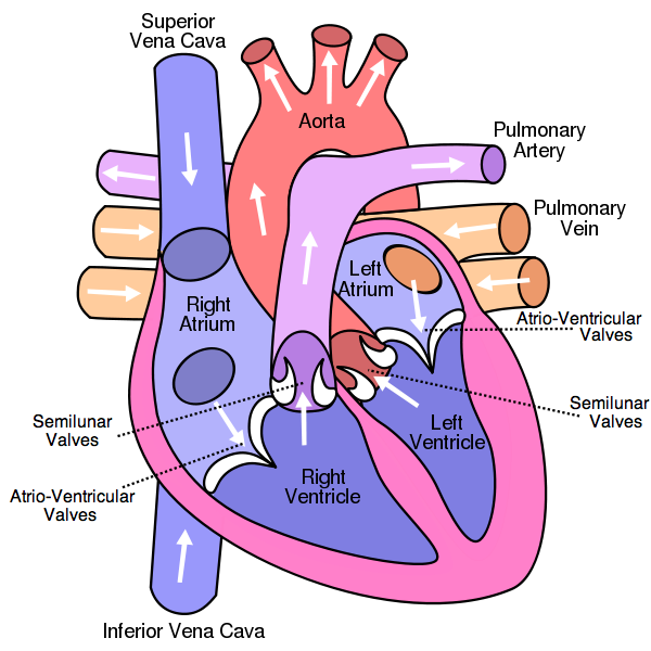
Heart Action: Blood Collection, Pumping, and Valve Function
The right atrium collects blood from the superior and inferior vena cava, and the left atrium collects blood from the pulmonary veins. This blood then flows into the right and left ventricles, which pump the blood into the arteries. The direction of blood flow is controlled by the atrioventricular valves and semilunar valves. When the atria contract, the blood flows through the open atrioventricular valves into the ventricles. At this stage, the semilunar valves are closed, so the ventricles fill with blood. The ventricles then contract, which causes a rise in pressure. This rise in pressure first causes the atrioventricular valves to close, preventing backflow of blood into the atria. Then, the semilunar valves open, allowing the expulsion of blood into the arteries. As this happens, the atria start to fill with blood again. The ventricles stop contracting, leading to a fall in pressure, which causes the semilunar valves to close, preventing backflow of blood from the arteries. When the ventricular pressure drops below the atrial pressure, the atrioventricular valves open again, and the cycle repeats.
Summary of Heart Action:
- Atria collect blood from veins.
- Atria contract, atrioventricular valves open.
- Blood is pumped into ventricles.
- Ventricles contract, atrioventricular valves close, and semilunar valves open.
- Blood is pumped into arteries; semilunar valves close.
- Cycle repeats.
Control of the Heartbeat: Myogenic Contraction and Regulation
Summary of Heartbeat Control:
- Heart muscle can contract by itself (myogenic muscle contraction).
- The pacemaker initiates contractions.
- One nerve carries messages from the brain to the pacemaker to speed up the heart's beating.
- Another nerve carries messages from the brain to the pacemaker to slow down the heart's beating.
- Epinephrine (adrenaline) signals the pacemaker to increase the heart's beating.
Blood Vessels: Structure and Function of Arteries, Veins, Capillaries
Arteries
- Thick outer layer of longitudinal collagen and elastic fibers prevents leaks and bulges.
- Thick wall withstands high pressure.
- Thick layers of circular elastic fibers and muscle fibers to pump blood.
- Narrow lumen to maintain high pressure.
Veins
- Thin layer with few circular elastic fibers and muscle fibers, as blood does not flow in pulses.
- Thin walls so that nearby muscles can help push blood towards the heart.
- Thin outer layer of longitudinal collagen and elastic fibers, as pressure is low.
- Wide lumen to accommodate the slow-flowing blood.
Capillaries
- Wall is one cell layer thick, so distance for diffusion is small.
- Pores allow plasma to leak out and form tissue fluid. Phagocytes can also pass through pores.
- Very narrow lumen so that many can fit in a small space.
Antibiotics: Effectiveness Against Bacteria, Not Viruses
Summary:
- Antibiotics block specific metabolic pathways in bacteria.
- Bacteria are very different from human cells, so human cells are not affected.
- Viruses require host cells to carry out metabolic processes for them, so antibiotics cannot be used to treat viruses.
- Harming the virus would harm the human cells.
First Line of Defense: Skin and Mucous Membranes
Skin
- Forms a physical barrier.
- Sebaceous glands secrete lactic acid and fatty acids.
Mucous Membranes
- Mucus contains lysozyme enzymes.
- Mucus can be sticky and trap pathogens.